The Carotid Sheath, the Styloid Process, and Eagle Syndrome: A Hidden Vagal Compression
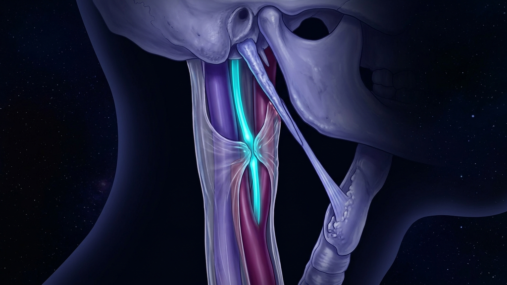
A patient feels a sharp throat pain that worsens when they turn their head, a fluttering heart, a sense that something is stuck when they swallow, and occasionally they nearly black out. Cardiology finds nothing structural; ENT finds nothing obvious. The problem is that the vagus nerve runs bundled with the carotid artery inside a single connective-tissue sleeve — and a bony spike from the base of the skull can sit close enough to irritate it on every movement. This is a hidden vagal compression, and its most defined form is Eagle syndrome.
The Bundle: What the Carotid Sheath Is
The carotid sheath (a tube of connective tissue running down each side of the neck that bundles three critical structures together) is the anatomical setup that makes this compression possible. Inside one sleeve travel:
- The common and internal carotid artery — carrying the majority of blood to the brain.
- The internal jugular vein — the dominant drainage route out of the cranium.
- The vagus nerve (cranial nerve X) — the parasympathetic master cable, running between the artery and vein for the length of the neck.
The consequence is intimacy: the vagus has no private channel. Anything that deforms the sheath — pressure, stretch, a pulsating artery pressed against a hard edge — is transmitted directly to the nerve. The baroreceptors of the carotid sinus, which sense blood pressure, sit at the same level. Irritate this region and you are pulling on both the autonomic sensor and the autonomic wire at once.
The Spike: The Styloid Process
Just behind and above the carotid sheath, a thin bony projection points down from the base of the skull: the styloid process (a slender spike of bone in front of the ear from which several throat muscles and a ligament hang). In most people it measures around 20-30 mm. But when it is abnormally long, or when the stylohyoid ligament (the cord tethering the styloid tip toward the hyoid bone) calcifies into rigid bone, the spike can extend deep into the neck and sit millimeters from the carotid sheath.
The numbers frame the problem. A styloid is generally considered elongated beyond about 30 mm, and elongation is found in roughly 4% of the general population on imaging. Yet only a small fraction — on the order of 4-10% of those with an elongated styloid — ever develop symptoms. Elongation is common; the collision with the neurovascular bundle is what makes it matter.
Eagle Syndrome: When the Spike Meets the Bundle
Eagle syndrome (symptoms caused by an elongated styloid process or a calcified stylohyoid ligament irritating nearby nerves and vessels) comes in two overlapping forms:
- The classic form irritates the glossopharyngeal nerve and throat tissues — throat pain, a foreign-body sensation on swallowing, ear pain, and pain that sharpens on turning the head.
- The vascular / neurovascular form is the one that masquerades as heart disease and dysautonomia. Here the elongated styloid presses on the carotid sheath itself, compressing the artery, vein, and vagus.
Why It Mimics a Heart Problem
When the spike loads the carotid sheath, the vagus and the carotid baroreceptors are provoked together, and the symptoms are autonomic:
- Palpitations and heart-rate swings — mechanical vagal irritation destabilizes the input to the sinoatrial node, the heart’s pacemaker.
- Syncope and near-syncope — pressure on the carotid sinus triggers an exaggerated baroreflex (the reflex that drops heart rate and blood pressure to defend against a perceived surge). A styloid pressing there can cause reflex fainting on head rotation, sometimes called styloid-carotid or carotid-sinus type Eagle syndrome.
- Throat and swallowing symptoms — the sensation of a lump, pain radiating to the ear, and discomfort that peaks with head turning.
- Positional provocation — because rotation swings the styloid toward the sheath, symptoms are reliably worse on turning the head to one side.
A patient with rotation-triggered palpitations and fainting who is worked up entirely as a cardiac case will keep coming back "normal," because the lesion is a bone spike two centimeters away from the heart’s nerve supply, not in the heart at all.
Locating and Confirming It
Like the other vagal choke points, this one has an address. The styloid tip lies deep between the angle of the jaw and the mastoid; in some patients an elongated, calcified styloid can be palpated in the tonsillar fossa at the back of the throat, and pressing it reproduces the pain. Confirmation is structural: a CT scan with 3D reconstruction is the reference standard, measuring styloid length and showing its relationship to the carotid sheath directly. Reproduction of symptoms on head rotation, plus an elongated styloid on imaging that tracks the neurovascular bundle, closes the loop.
What This Means for Practitioners
- Put Eagle syndrome on the list for unexplained throat pain, ear pain, palpitations, or rotation-triggered near-syncope — especially when cardiac and standard ENT workups are unremarkable.
- Test with rotation. Symptoms that reproduce on turning the head toward the affected side point at a moving styloid, not a fixed cardiac lesion.
- Image the bone, measure the length. A styloid beyond roughly 30 mm that projects toward the carotid sheath is the finding that converts a vague symptom into a mechanical diagnosis.
- Frame it correctly for the patient. These are not psychosomatic palpitations. A calcified ligament and an overgrown spike are irritating the vagus and the carotid baroreceptor bundle — a locatable, treatable structural problem.
The carotid sheath forces the vagus to share a tube with a pulsating artery and a major vein, and the styloid process is a bony blade poised beside that tube. Eagle syndrome is what happens when the blade grows long enough to reach. Name the structure, image the spike, and a "heart condition of unknown cause" resolves into anatomy you can point to.
Reference: European Archives of Oto-Rhino-Laryngology, elongated styloid process and Eagle syndrome (2022).