The Anxiety–Mitochondria Connection: Calming Cells to Calm the Mind
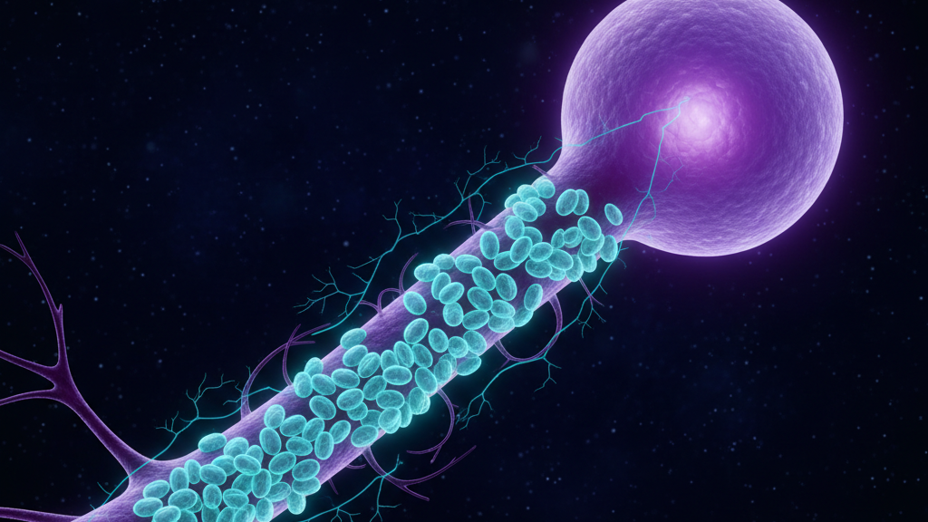
For decades anxiety has been framed as a neurotransmitter problem — too much glutamate, too little GABA, dysregulated serotonin. That model is not wrong. It is incomplete. The latest mitochondrial psychiatry literature suggests something more fundamental: a brain that cannot reliably make enough ATP defaults to a state of vigilance.
Anxiety as an Energy Story
The amygdala, the prefrontal cortex, and the locus coeruleus are among the most metabolically demanding tissues in the body. When mitochondrial output drops — from chronic stress, sleep loss, infection, micronutrient depletion, or genetic vulnerability — these circuits do not fail uniformly. The inhibitory networks fail first. The result is a brain biased toward threat detection, with reduced top-down regulation. That phenomenology is anxiety.
What the Patient Reports
The clinical picture is consistent: an anxious patient often arrives with cold extremities, low HRV, post-exertional symptom flares, mid-afternoon energy crashes, and sleep that is long but unrefreshing. These are not coincidental. They are markers of impaired bioenergetics.
The Cellular Calm Protocol
I structure intervention around restoring mitochondrial capacity, not just dampening symptoms.
- Magnesium glycinate — supports ATP synthesis and is a cofactor for several enzymes in the citric acid cycle.
- CoQ10 (ubiquinol form) — directly supports the electron transport chain.
- Omega-3 fatty acids — stabilize mitochondrial membranes and reduce neuroinflammation.
- Slow exhalation breathing — biases the autonomic nervous system toward parasympathetic dominance, reducing the metabolic cost of vigilance.
- Morning sunlight — entrains circadian rhythm, which gates mitochondrial repair cycles.
Where Ultrasound Neuromodulation Comes In
Low-intensity focused ultrasound applied to the right amygdala has shown reductions in subjective anxiety in controlled trials, with a mechanism that appears to involve reduced amygdalar metabolic activity. This is consistent with the broader thesis: dial down the metabolic demand of the threat circuit, and the system has the room to recover.
The Microtubule Footnote
Microtubules inside neurons are not just structural scaffolding; they appear to participate in intracellular signaling and may play a role in synaptic plasticity. Anesthetics that bind microtubule hydrophobic pockets reliably reduce anxiety. This is suggestive — not conclusive — that microtubule dynamics may be one of several underappreciated levers in anxiety physiology.
Bottom line: If a patient is "trying everything" for anxiety and not improving, run the bioenergetic workup. Often the missing variable is in the cell, not in the cortex.
References
- Picard M, McEwen BS, Epel ES. "An energetic view of stress: Focus on mitochondria." Frontiers in Neuroendocrinology, 2018;49:72-85.
- Hollis F et al. "Mitochondrial function in the brain links anxiety with social subordination." PNAS, 2015;112(50):15486-15491.
- Mahoney JJ et al. "Low-intensity focused ultrasound targeting the bilateral amygdala in adults." Frontiers in Human Neuroscience, 2023;17:1196696.