Vagal Axon Degeneration in Long COVID: What the Biopsies Suggest
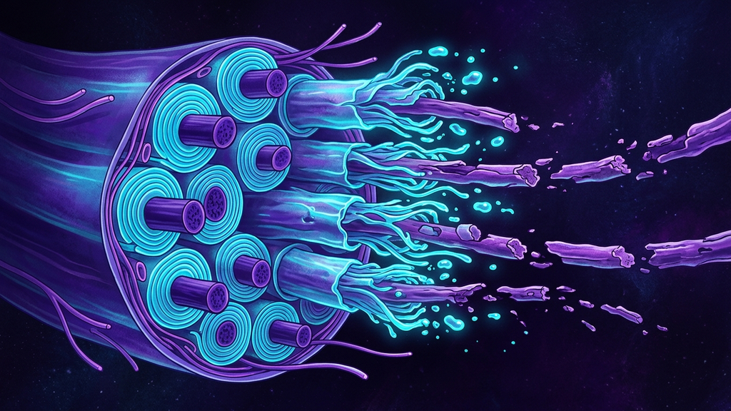
A patient recovers from a viral infection but never recovers their energy. Their heart races when they stand. Their gut slows. Their thinking blurs. For a long time the reflexive verdict was psychological: deconditioning, anxiety, stress. But when researchers began looking at nerve tissue directly, they found something the psychological model cannot explain: signs of physical injury to the nerve fibers themselves. This is emerging, still-limited evidence, and it must be read carefully. But it reframes a dismissed symptom cluster as a problem of damaged wiring, and the wiring in question runs through the vagus nerve.
What "Degeneration" Actually Means
Nerves are cables. The signal travels down the axon (the long conducting fiber of a nerve cell), and many axons are wrapped in myelin (the fatty insulating sheath that lets signals travel fast and cleanly). Injury shows up as two distinct lesions.
- Demyelination (loss of the myelin insulation) leaves the axon intact but stripped, so signals slow, scatter, or fail intermittently.
- Axonal degeneration is worse: the conducting fiber itself dies back. When an axon is cut off or fatally injured, the segment beyond the injury self-destructs in a stereotyped process called Wallerian degeneration (the orderly disintegration of the nerve fiber downstream of an injury). This is not the nerve being tired. It is the nerve dismantling itself.
Where the Vagus Comes In
The vagus is unusually vulnerable to this because so much of it is thin and small-calibre. Alongside its large myelinated fibers it carries vast numbers of tiny unmyelinated ones, the same class implicated in small-fiber neuropathy (damage to the smallest sensory and autonomic nerve fibers, diagnosable by skin biopsy). And roughly 80% of vagal fibers are afferent (carrying sensory information from the organs up to the brain). If those afferents degrade, the brain loses its readout of the body, of heart, lungs, and gut, which is precisely the picture patients describe.
Post-viral injury to these fibers has a plausible route. Some viruses show neurotropism, inflammation can damage the tiny vessels that feed nerves, and autoimmune cross-reactivity can attack myelin. Any of these can produce the two lesions above in autonomic and vagal fibers.
What the Evidence Actually Shows
Honesty about strength of evidence matters here, because the claim is consequential.
- Multiple small skin-biopsy studies of long-COVID cohorts have reported small-fiber neuropathy in a substantial subset, some series finding evidence of small-fiber pathology in roughly 60% of biopsied patients with post-COVID autonomic symptoms. These are small cohorts, not population studies.
- Imaging work using neck ultrasound has reported reduced vagus nerve cross-sectional diameter in some post-COVID patients versus controls, a structural, not psychological, finding.
- Autopsy and early biopsy reports have described axonal degeneration and inflammatory infiltrates in vagal and brainstem tissue in some post-viral cases, though these are limited case-level observations, not confirmed prevalence.
- Given that ~80% of vagal fibers are afferent, even partial afferent loss can disproportionately degrade interoceptive signaling.
The right summary is measured: the biopsy and imaging evidence is emerging and limited, drawn from small cohorts and case series. It does not yet prove that every fatigued post-viral patient has a degenerating vagus. But it is enough to say the structural injury hypothesis is real, testable, and no longer dismissible.
How the Two Lesions Present Differently
The distinction between demyelination and axonal degeneration is not academic; it predicts the recovery curve. A demyelinated but intact fiber can, in principle, be re-insulated on a timescale of weeks, so a patient whose signal is merely slowed has a more favorable outlook. A fiber that has undergone Wallerian degeneration must be regrown from the injury site outward, which is slower and less certain. Two patients with identical symptoms can therefore sit on very different trajectories depending on which lesion dominates, and that is exactly why tissue-level and imaging evidence matters rather than symptom scoring alone.
The clustering also makes sense once you accept the structural model. Post-viral patients tend to present not with one isolated complaint but with a constellation, an orthostatic heart-rate surge, sluggish digestion, temperature dysregulation, and blunted body-awareness, precisely because a single injured nerve carries fibers to many of those systems at once. The symptoms cluster because the wiring is shared.
Why the Reframe Matters
If a vagal afferent has undergone Wallerian degeneration, no amount of reassurance will regrow it. The symptom is not a belief; it is a broken cable. This changes the clinical posture entirely. This is not anxiety producing a racing heart, it is a damaged sensor failing to report the body's state, and an autonomic system running without its feedback. Validating that mechanically, rather than psychologizing it, is itself part of good care.
It also reframes the timeline. Structural nerve injury does not resolve on the schedule of a mood; it follows the slow biology of nerve repair, which is measured in weeks and months, not days. A patient who is not "better by now" is not failing to try, their axons are on their own clock.
The Takeaway for Practitioners
Treat unexplained post-viral dysautonomia as a possible structural neuropathy until proven otherwise. That means taking objective signs seriously, such as an exaggerated heart-rate rise on standing, gut dysmotility, and blunted interoception, and recognizing them as candidate readouts of vagal fiber loss rather than character flaws. The vagus can be injured. Increasingly, the tissue evidence suggests that in some long-COVID patients, it has been.
Reference: skin-biopsy and neuroimaging studies of post-COVID small-fiber and autonomic neuropathy, Neurology and related journals (2021-2024).