Ultrasound Neuromodulation — Mechanism of Action at the Cellular Level
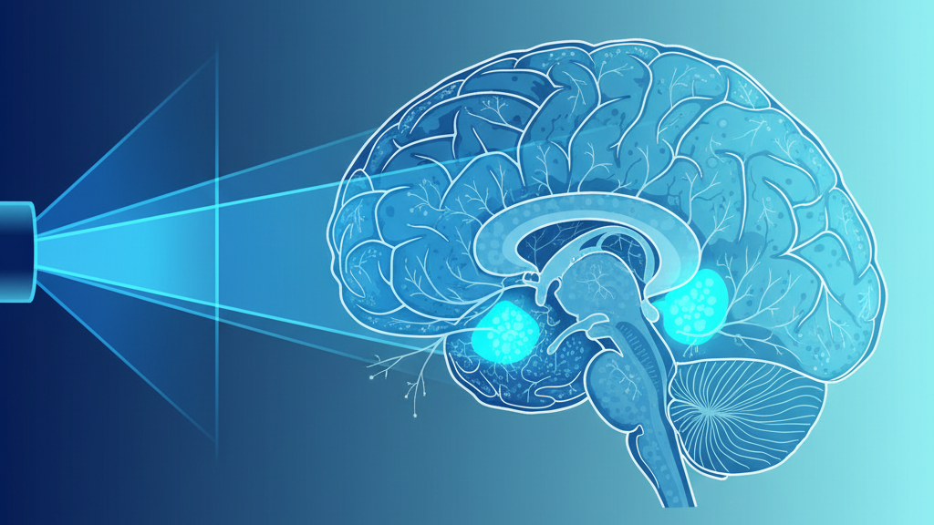
Low-intensity focused ultrasound (LIFU) has emerged in the last decade as the most promising non-invasive neuromodulation tool we have. It reaches deep brain structures with millimeter precision, requires no surgery, leaves no permanent change, and can both excite and inhibit neurons depending on the pulse parameters. The clinical results are increasingly hard to dismiss. The mechanism, however, remains genuinely open — and the answer matters more than it might first appear.
What LIFU Actually Does to Neurons
The textbook accounts of ultrasound neuromodulation invoke four candidate mechanisms, often combined:
- Acoustic radiation force. A travelling pressure wave exerts a steady push on tissue, mechanically deforming cell membranes.
- Cavitation. At higher intensities, microbubbles in the tissue oscillate or collapse, producing localized shear forces.
- Mechanosensitive ion channels. Membrane proteins like Piezo1, Piezo2, and several TRP-family channels open in response to mechanical strain.
- Mild local heating. Even sub-thermal protocols produce a small temperature rise that can change membrane properties.
Each of these is real, and each contributes. Together, however, they fail to explain a central observation: LIFU's effects are unusually precise, reversible, and parameter-sensitive. Small changes in pulse repetition frequency or duty cycle flip the response from excitation to inhibition. Whatever LIFU is doing, it is not just shaking the tissue.
Mechanosensitive Channels — Necessary, Not Sufficient
Piezo1 and Piezo2 are the best-characterized mechanosensitive channels in the nervous system. They open when the membrane is stretched and let calcium and sodium in. Genetic knockouts of Piezo1 sharply reduce the neuronal response to LIFU, which establishes them as part of the pathway.
But the response in Piezo-knockout animals is reduced, not abolished. Some other transduction mechanism is also at work. The leading candidate, increasingly, is the cytoskeleton itself.
The Cytoskeletal Hypothesis
Microtubules are mechanically active structures. They generate force during cell division and during the transport of organelles. They are also piezoelectric: mechanical strain produces electrical polarization, and electrical fields produce mechanical strain. This makes them inherently responsive to ultrasound, which is precisely a mechanical pressure wave.
Three lines of evidence point to microtubules as a primary LIFU target:
- Resonance match. Microtubule lattices have intrinsic vibrational modes spanning the megahertz to low-gigahertz range. The most therapeutically effective LIFU frequencies (250–650 kHz) drive harmonics that couple efficiently into these modes through bundled axons.
- Anesthetic overlap. Anesthetics that abolish consciousness bind directly to tubulin. LIFU appears, in some protocols, to reverse sedation. This is exactly what you would expect if the two interventions push microtubule dynamics in opposite directions.
- Therapeutic targets. The clinical conditions where LIFU shows the most promise — Alzheimer's, depression, PTSD, traumatic brain injury — all involve documented cytoskeletal disruption.
"The mechanism of ultrasound neuromodulation remains incompletely understood. The mechanosensitive ion channel hypothesis is necessary but not sufficient. A complete account will likely involve direct mechanical coupling to subcellular structures including the cytoskeleton." — Blackmore et al., Ultrasound in Medicine & Biology, 2019
The Quantum-Coherence Permutation
If microtubules support quantum-coherent states (the strong form of the Orch-OR hypothesis), then LIFU's mechanism becomes more interesting still. Acoustic phonons at the right frequency would not merely deform the lattice — they would reset or extend coherence times by driving the system through specific vibrational modes. In this view, LIFU works because it tunes the cellular substrate of consciousness directly, not because it tickles ion channels.
This is a strong claim and currently lacks direct experimental confirmation. But it is not idle speculation: it predicts that LIFU's therapeutic effects should depend critically on frequency in a way that simple mechanical models do not predict. That prediction is testable.
Why the Mechanism Question Matters
Two competing ideas about how LIFU works produce two different roadmaps for the next decade of clinical development:
- If LIFU acts primarily through Piezo channels, the way to improve it is to identify cell populations enriched in those channels and target them directly.
- If LIFU acts primarily through cytoskeletal resonance, the way to improve it is to match the frequency to the structural dimensions of the target tissue — and to pair LIFU with interventions that support cytoskeletal health.
These lead to very different devices, very different protocols, and very different patient selection criteria. The mechanism question is not academic. It is the next bottleneck.
Further Reading
Blackmore J., Shrivastava S., Sallet J., Butler C.R., & Cleveland R.O. (2019). Ultrasound Neuromodulation: A Review of Results, Mechanisms and Safety. Ultrasound in Medicine & Biology, 45(6), 1509–1536. doi:10.1016/j.ultrasmedbio.2018.11.017