MCAS, the Vagus Nerve, and the Cholinergic Anti-Inflammatory Reflex
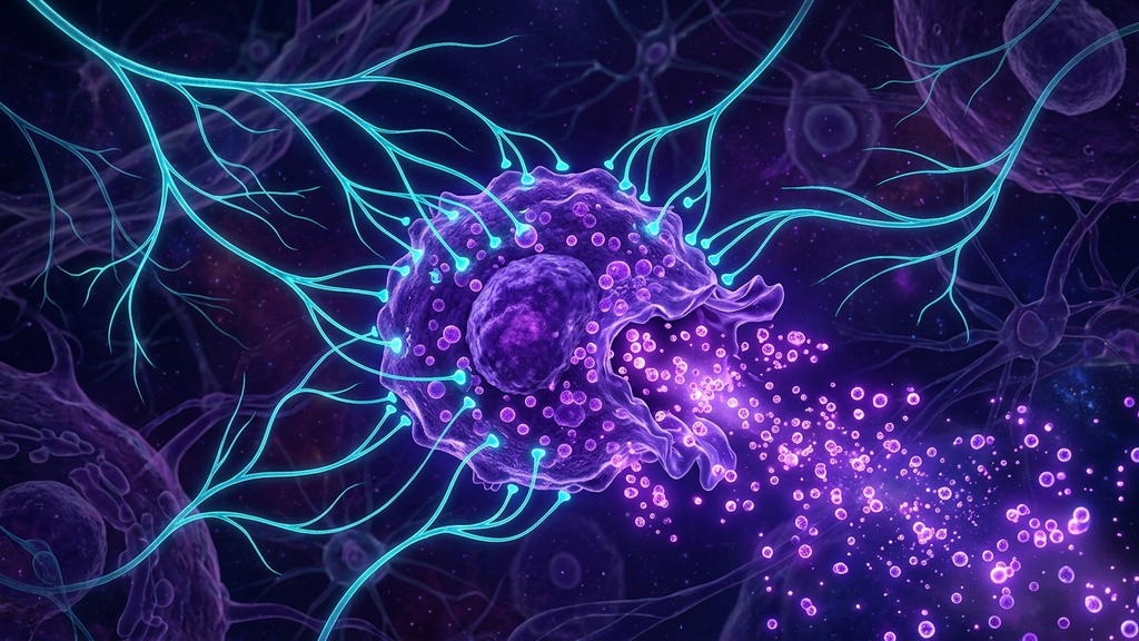
Mast Cell Activation Syndrome has emerged from the fringe to the center of complex chronic illness. The reason is straightforward: mast cells sit at the intersection of immunity, autonomic signaling, and connective tissue biology, and when they fire inappropriately, they produce the polysymptomatic pictures that define hard-to-categorize patients — flushing, hives, palpitations, GI distress, brain fog, anxiety, food sensitivities, and unpredictable flares.
What Mast Cells Actually Do
Mast cells are tissue-resident innate immune cells embedded in connective tissue, especially around blood vessels and nerve endings. They store granules packed with histamine, tryptase, heparin, prostaglandins, and over 200 mediators. When activated — by allergens, infection, mechanical pressure, temperature shifts, hormonal changes, or stress — they release these mediators into surrounding tissue. The result is the familiar inflammatory cascade.
The Vagal Connection
Here is the underappreciated piece: mast cells are richly innervated by the vagus nerve, and acetylcholine — the vagal neurotransmitter — modulates mast cell activation through nicotinic and muscarinic receptors. The most consistent evidence supports an inhibitory effect at the α7 nicotinic acetylcholine receptor (α7nAChR). When vagal tone is high, the cholinergic anti-inflammatory pathway suppresses mast cell degranulation. When vagal tone is low, mast cells release more readily.
"Activation of nicotinic receptors inhibited vagus-stimulated histamine release in a murine food allergy model. The cholinergic anti-inflammatory pathway provides a real, modulatable brake on mast cell activity."
Why MCAS Patients Often Have Low Vagal Tone
The relationship is bidirectional, which is why these patients spiral. Mast cell mediators (histamine, tryptase, prostaglandins) directly activate sympathetic afferents and depress vagal tone. Low vagal tone removes the brake on mast cells. Mast cells fire more easily. The system runs hotter. Patients describe the experience as their nervous system being "set on fire."
The Diagnostic Picture
Formal MCAS diagnosis requires elevated mast cell mediators on lab testing, but in practice the clinical phenotype is recognizable:
- Reactions to multiple unrelated triggers (foods, scents, heat, stress, alcohol, exercise).
- Flushing, hives, dermatographism, or unexplained itch.
- Episodic palpitations, GI distress, and brain fog with no consistent pattern.
- Frequent overlap with POTS and hypermobility (the EDS-POTS-MCAS triad).
- Reaction patterns that worsen with stress and sleep deprivation — the autonomic-immune link.
The Treatment Framework
Stabilize the mast cell
- H1 antihistamine (e.g., cetirizine, fexofenadine) — twice-daily dosing.
- H2 antihistamine (e.g., famotidine) — important and often missed.
- Mast cell stabilizer — cromolyn sodium (oral), quercetin, luteolin.
- Vitamin C (acts as a histamine cofactor for clearance).
Restore the vagal brake
- Slow nasal breathing protocols — measurable HRV improvement within days.
- Transcutaneous auricular vagus nerve stimulation (taVNS) — targets the cholinergic anti-inflammatory pathway directly.
- HRV biofeedback — daily training over weeks rebuilds parasympathetic reserve.
- Cold exposure (titrated) — a vagal afferent stimulus, but caution in MCAS where cold itself can be a trigger.
Reduce overall inflammatory load
- Investigate environmental triggers — mold exposure is a major underrecognized driver.
- Address gut dysbiosis and barrier dysfunction.
- Identify and remove hidden chronic infections (Lyme, EBV, HHV-6).
- Hormonal stabilization — estrogen spikes are mast cell triggers in many women.
What "Mast Cell-Friendly Living" Actually Looks Like
Patients often arrive with elaborate restriction lists. The most clinically useful structure is broader: reduce total inflammatory load (food triggers, environmental triggers, sleep deprivation, sympathetic dominance), restore the vagal brake, and stabilize the cell pharmacologically while the system rebuilds. Patients who try to control MCAS through avoidance alone usually end up with shrinking diets and growing reactivity.
Clinical takeaway: MCAS responds to the vagal-anti-inflammatory lever. Stabilize the cell pharmacologically, restore vagal tone, reduce inflammatory inputs, and the reactivity ladder slowly resets. Avoidance alone fails; the brake has to be rebuilt.
References
- Theoharides TC et al. "Mast cell activation syndrome: a state-of-the-art review." Journal of Allergy and Clinical Immunology, 2024;153(6):1655-1670.
- Forsythe P. "Mast cells in neuroimmune interactions." Trends in Neurosciences, 2019;42(1):43-55.
- van Westerloo DJ et al. "The vagus nerve and nicotinic receptors modulate experimental pancreatitis severity in mice." Gastroenterology, 2006;130(6):1822-1830.
- Tracey KJ. "The inflammatory reflex." Nature, 2002;420:853-859.
- Caringi M et al. "Mast cells in the autonomic nervous system and potential role in disorders with dysautonomia and neuroinflammation." Annals of Allergy, Asthma & Immunology, 2024;132(4):440-449.