The Jugular Foramen Bottleneck: The Skull Opening Where the Vagus Gets Choked
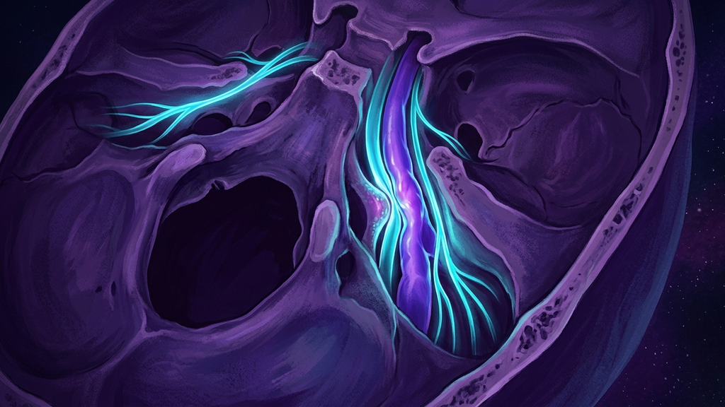
A patient reports pulsatile tinnitus, positional dizziness, a heart that races for no reason, and a fog that never fully lifts. Every scan comes back "normal," so the file is stamped anxiety. But there is a specific place in the skull where four critical structures are forced through one narrow bony doorway — and when that doorway is crowded, every one of those symptoms follows mechanically. The place has a name: the jugular foramen. Call the phenomenon the jugular foramen bottleneck.
One Doorway, Four Occupants
The jugular foramen (a paired opening at the base of the skull where nerves and a major vein exit the cranium) is one of the most crowded passages in the human body. Through a gap that in most adults measures only about 10-15 mm at its widest, four structures must pass simultaneously:
- The vagus nerve (cranial nerve X) — the primary parasympathetic highway to the heart, lungs, and gut, carrying roughly 80% of its fibers as afferent (sensory) traffic back toward the brain.
- The glossopharyngeal nerve (cranial nerve IX) — which relays the baroreflex and controls part of the throat and taste.
- The accessory nerve (cranial nerve XI) — which drives the sternocleidomastoid and trapezius muscles of the neck.
- The internal jugular vein — the dominant drainage route returning blood and cerebrospinal fluid from the brain, handling the majority of cranial venous outflow on its side.
Nerve tissue and venous tissue sharing one rigid bony tunnel is an engineering compromise. When the tunnel narrows, or when the vein inside it swells, there is nowhere for the pressure to go.
How the Bottleneck Chokes
Three mechanisms crowd the foramen, often in combination:
Bony stenosis
The foramen itself can be congenitally small or narrowed by bony overgrowth. Imaging series have found jugular foramen stenosis (structural narrowing of the opening) in a meaningful subset of patients presenting with unexplained pulsatile tinnitus, with degrees of luminal narrowing exceeding 50% in symptomatic cases. A tunnel that is 30-50% narrower leaves far less tolerance for any additional swelling.
Venous congestion
Because the internal jugular vein shares the space, anything that engorges the vein presses directly on the adjacent nerves. Internal jugular vein compression or stenosis has been documented in a large fraction of patients with chronic cerebral venous outflow complaints, and the resulting back-pressure raises intracranial venous volume. That congestion is a leading structural explanation for pulsatile tinnitus — the patient literally hears turbulent flow next to the cochlea.
Dynamic compression from above and beside
The foramen sits directly beneath the C1 vertebra and behind the styloid process. Rotation of the head, an elongated styloid, or instability at the top of the neck can dynamically pinch the vein and nerves as they emerge. This is why symptoms are so often positional — worse when lying on one side, turning the head, or looking up.
Why It Produces That Exact Symptom Cluster
The bottleneck is elegant in its cruelty because each occupant maps onto a specific complaint:
- Vagus (CN X) irritation destabilizes autonomic tone — palpitations, a heart rate that jumps, nausea, air hunger, and the surges of a body that cannot settle. This is dysautonomia produced at the wire, not in the mind.
- Glossopharyngeal (CN IX) involvement distorts the baroreflex (the blood-pressure sensing loop that keeps you from fainting when you stand), feeding lightheadedness and blood-pressure swings.
- Internal jugular congestion raises intracranial pressure and slows the clearance of metabolic waste from the brain — the mechanical substrate of brain fog, pressure headaches, and pulsatile tinnitus.
- Accessory (CN XI) irritation loads the neck muscles, producing the stubborn one-sided neck and shoulder tightness that so many of these patients carry.
Four symptoms that look like four unrelated problems — or like a psychiatric label — resolve into one anatomical fact when you locate them at the foramen.
You Can Locate This Spot
This is not an abstract deep-brain structure. The jugular foramen sits roughly a fingertip-width behind the angle of the jaw and just below the ear canal, deep to the mastoid tip. The vagus and its neighbors are traveling within about 1-2 cm of the skin surface there before they descend into the neck. For a practitioner, palpating that region — and noting reproduction of dizziness, throat sensation, or heart-rate change with gentle positional pressure or head rotation — is a legitimate bedside localization. The symptom has an address.
What This Means for Practitioners
The jugular foramen bottleneck reframes a frustrating class of patients. When someone presents with the tetrad of pulsatile tinnitus, positional dizziness, autonomic instability, and unilateral neck tension — especially if standard brain MRI is unremarkable — the skull base deserves dedicated attention:
- Consider positional or dynamic imaging and dedicated venous studies. A vein that looks patent lying flat can collapse on rotation; static supine imaging misses the mechanism by design.
- Screen for the neighbors — an elongated styloid process and upper-cervical instability both narrow this doorway and are addressable.
- Treat the venous side seriously. Because congestion is often the driver, interventions that reduce venous back-pressure or upper-cervical mechanical load target the actual bottleneck rather than sedating the patient.
The reframe matters most for the patient. A young person told for years that their racing heart and ringing ears are anxiety is being handed a psychological story for a structural problem. The vagus is not neurotic. It is being compressed in a 12-millimeter tunnel it shares with three other structures and the brain’s main drain. Name the bottleneck, image it dynamically, and the invisible becomes locatable.
Reference: Otology & Neurotology, jugular foramen stenosis and pulsatile tinnitus (2023).