The Spleen-Brain Axis: How the Vagus Commands the Body's Inflammation Thermostat
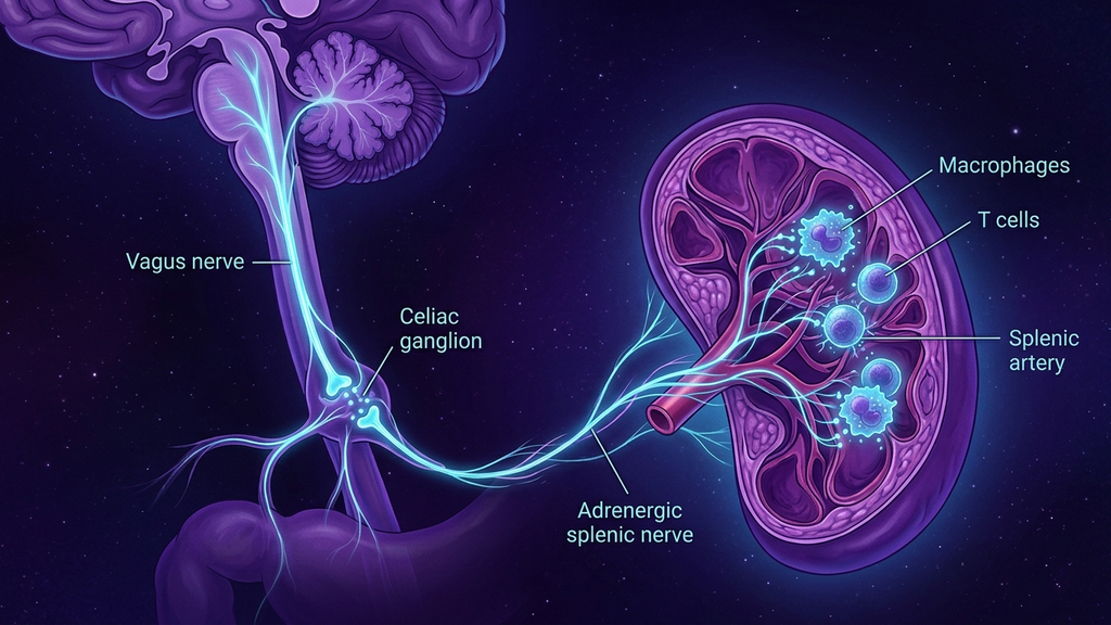
Immunologists once believed inflammation was a purely local, chemical affair, cells shouting cytokines at one another until something told them to stop. In 2000, Kevin Tracey mapped the something. He showed the brain and immune system are wired together by a physical neural circuit that measures inflammation and, within seconds, issues a command to suppress it. The organ that receives that command is not the liver or the gut. It is the spleen. This is the spleen-brain axis, and it may be the single most important brake on the chronic inflammation that underlies modern disease.
The Circuit, Not the Chemistry
The system Tracey named is the inflammatory reflex (a hardwired neural loop in which the vagus nerve both senses and controls inflammation). Like any reflex, it has a sensory arm and a motor arm.
The sensory, or afferent (carrying signals from the body up to the brain), arm does the reading. Roughly 80% of the fibers in the vagus nerve are afferent, and many terminate near sites where the body samples its own internal chemistry. When cytokines such as TNF and IL-1 rise, these fibers fire and carry the message to the brainstem. The brain, in effect, gets a real-time readout of the body's inflammatory temperature.
The motor, or efferent, arm sends the correction back down. And here is where the anatomy becomes surprising: the vagus does not travel all the way to the spleen. Instead it relays through the celiac ganglion (a cluster of nerve cell bodies in the upper abdomen that acts as a relay station), which gives rise to the splenic nerve (the adrenergic nerve that runs alongside the splenic artery into the spleen). It is this splenic nerve that carries the final signal into the organ.
Why the Spleen Is the Hub
The spleen is not a passive filter. It is the largest secondary immune organ in the body, and at any moment it holds a substantial reservoir of the body's monocytes and lymphocytes, a garrison of inflammatory cells waiting for orders. Controlling the spleen means controlling a huge fraction of systemic cytokine output from a single point. In network terms, it is a bottleneck the nervous system can exploit.
The mechanism inside the organ is elegant. The splenic nerve releases noradrenaline. That noradrenaline acts on a specialized population of T cells that express choline acetyltransferase, the ChAT+ T cells (immune cells capable of manufacturing the neurotransmitter acetylcholine). Prompted by the nerve, these T cells release acetylcholine, which binds alpha-7 nicotinic receptors on neighboring macrophages. That binding is the off-switch: it suppresses NF-kB signaling and throttles the release of TNF and IL-6. This nerve-to-immune handoff is the cholinergic anti-inflammatory pathway (the acetylcholine-mediated final step that silences macrophage cytokine release), and the spleen is where it physically happens.
The Afferent Alarm
It is worth dwelling on the sensory half, because it is the part most people miss. The vagus is not merely a control cable running out to the organs; it is overwhelmingly an incoming line. Those afferent fibers are studded with receptors that respond to the molecular signature of infection and injury, so the nervous system does not have to wait for the immune system to make it feel ill. It detects rising cytokines directly and can begin adjusting behavior and physiology before inflammation escalates. When this afferent alarm is intact, the brain and immune system negotiate in real time. When it is degraded, the loop runs open, and the first sign is often a mismatch between how inflamed a patient measurably is and how poorly the body is regulating that inflammation.
The Numbers
- Approximately 80% of vagal fibers are afferent, so the circuit is built more for listening than for commanding.
- In animal models, electrical stimulation of the vagus or splenic nerve can cut serum TNF by roughly 70% during an inflammatory challenge.
- Cutting the splenic nerve, or removing the spleen entirely, largely abolishes the anti-inflammatory effect of vagus nerve stimulation, direct proof the spleen is the required endpoint.
- TNF and IL-6 are the two most commonly measured readouts of the pathway; both fall when the axis fires and climb when it is severed.
What Frays the Wire
The spleen-brain axis fails at the level of signal strength. Low vagal tone, measurable indirectly through reduced heart-rate variability, means a weaker efferent command reaching the celiac ganglion and splenic nerve. The garrison in the spleen stops receiving its stand-down order, and macrophages default to their inflammatory setting. This is one mechanistic reason chronic low vagal tone travels alongside conditions of runaway inflammation: cardiovascular disease, autoimmunity, metabolic syndrome, and post-viral inflammatory states.
Crucially, the failure can be structural or functional. A vagus that is compressed, demyelinated, or degenerated cannot relay a clean signal no matter how healthy the spleen is. The axis is only as strong as its weakest segment, from brainstem to splenic artery.
What This Means for Practitioners
The clinical reframe is this: systemic inflammation is not only an immune problem to be suppressed with drugs, it is also a neural regulation problem. A patient with stubborn inflammatory markers and low heart-rate variability may not have an overactive immune system so much as an underpowered brake.
That opens a second lever. Anything that genuinely raises vagal efferent output, such as paced breathing, cold exposure, and neuromodulation approaches that target vagal branches, is mechanistically an attempt to restore command over the splenic hub. The goal is not to sedate the immune system but to reconnect it to the circuit that was always meant to govern it. The spleen-brain axis is the body's inflammation thermostat, and the vagus is the wire that sets the dial.
Reference: Tracey, "The inflammatory reflex," Nature (2002); and subsequent work on splenic nerve signaling, Journal of Experimental Medicine (2011).