The Hepatic Vagal Branch: The Liver's Direct Line to the Brain for Glucose and Energy
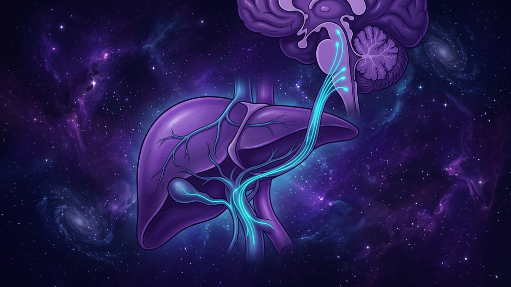
Ask most people how the brain knows the body's blood sugar, and they will describe glucose sensors in the brain itself, sampling the blood as it passes. That is real, but it is late information — by the time glucose has reached the brain, the meal is long absorbed. The body has a faster, more forward-positioned reporter: a slender branch of the vagus nerve that wraps the liver and the vessel feeding it, reading the incoming fuel supply at the point of entry and telegraphing it straight to the brainstem. This is the hepatic vagal branch, and it is one of the most anatomically specific and clinically overlooked nodes in the liver-brain axis that governs glucose and energy.
The anatomy: a dedicated line off the anterior trunk
As the vagus nerve descends through the chest and passes the esophagus, its fibers regroup into two trunks. From the anterior vagal trunk, a distinct hepatic branch (a dedicated nerve twig running to the liver) peels off and travels within the lesser omentum to reach the liver's hilum, where it distributes along the portal vein and the hepatic artery. It is small, but it is a direct, private channel between one metabolic organ and the brainstem.
Crucially, this branch is dominated by afferent fibers — sensory fibers carrying information from the liver up to the brain, rather than commands down to it. That fits the vagus as a whole, where roughly 80 percent of all fibers are afferent. The hepatic branch is therefore best understood not as a control cable but as a sensor lead: its main job is to report, and the report lands in the nucleus tractus solitarius (the brainstem hub that receives visceral sensory input) and from there reaches the hypothalamus, the brain's metabolic command center.
Portal glucose sensing: reading the fuel at the door
The liver sits in a privileged position. Everything absorbed from the intestine — glucose, amino acids, the products of digestion — is funneled first through the portal vein (the large vessel carrying nutrient-rich blood from the gut to the liver) before it reaches the general circulation. Embedded in and around that portal system are portal glucose sensors, specialized detectors that read the glucose concentration of this incoming stream.
These sensors report through the hepatic vagal branch. That gives the brain something the systemic circulation cannot: an early, upstream reading of nutrient inflow, taken before the glucose has diluted into the whole bloodstream. It is the difference between a lookout at the harbor mouth and a clerk counting ships after they have already docked and dispersed. The portal-vagal sensor tells the brain a meal is arriving and roughly how large, allowing pre-emptive adjustment of insulin dynamics, glucose uptake, and — importantly — the sense of having eaten.
Falling glucose speaks even louder than rising glucose
The hepatic afferent line is arguably most important not when glucose is high but when it is falling. The portal sensors are exquisitely tuned to a drop in glucose, and a decline detected here is one of the earliest triggers of the counter-regulatory response — the coordinated release of glucagon and adrenaline that mobilizes stored sugar to defend the brain's supply.
This is why the hepatic branch matters for anyone with reactive, shaky, adrenaline-flavored glucose symptoms. The portal-vagal sensor is often the first alarm in the chain. It detects the downslope of a post-meal glucose curve early and hard, and it can help trigger the sympathetic surge — the palpitations, the tremor, the wave of dread — well before a fingerstick would read anything alarming. The symptom is not the brain overreacting to nothing. It is the brain acting faithfully on an early warning delivered by a nerve doing exactly its job.
Appetite, satiety, and the liver's vote on hunger
The hepatic vagal branch is also a genuine appetite signal. Metabolic states in the liver — the availability of glucose and the oxidation of fats — are sensed and relayed upward, contributing to the brain's decision to start or stop eating. In animal models, cutting the hepatic vagal branch alters food intake and blunts the satiety normally produced by nutrients arriving at the liver, which pins the effect on this specific nerve rather than on hormones alone.
The practical reading is that satiety is not purely a stomach-stretch or a gut-hormone phenomenon. The liver casts a vote, and it casts it through the vagus. When hepatic afferent signaling is degraded — by chronic metabolic stress, by fatty-liver change, or by the general vagal blunting seen in dysautonomia — the brain loses part of its early nutrient-inflow and satiety information and must rely on later, coarser signals. Appetite regulation gets noisier, and the fine timing of "enough" is lost.
The hepatic branch, in numbers
- Roughly 80 percent of vagal fibers are afferent, and the hepatic branch is overwhelmingly a sensory, reporting line.
- All portal blood arrives first at the liver — essentially 100 percent of gut-absorbed nutrients pass this checkpoint before entering the general circulation, making it the earliest possible sampling site.
- Portal sensors respond to falling glucose ahead of systemic sensors, positioning the hepatic branch among the first triggers of counter-regulation rather than a late responder.
- Selective hepatic branch section in animal studies measurably changes food intake and nutrient-driven satiety, isolating this one twig as functionally significant.
The liver-brain axis as a control loop
Put together, the hepatic vagal branch anchors a true liver-brain axis: portal sensors read incoming fuel and hepatic metabolic state, the afferent hepatic branch relays that reading to the nucleus tractus solitarius, the brain integrates it in the hypothalamus, and the response comes back as adjusted insulin dynamics, glucose handling, and appetite. It is a closed loop, and the hepatic branch is its sensory limb. Interoception (the brain's perception of the body's internal state) includes this hepatic glucose readout, even though we never consciously feel it — until the loop misfires and the misfire surfaces as a symptom.
What this means for practitioners
The hepatic branch is not a target you stimulate with a device in a coach's office, but understanding it sharpens how you interpret and manage metabolic-autonomic symptoms.
- Reframe reactive glucose symptoms mechanically. When a patient with normal labs describes shaky, panicky post-meal crashes, the early adrenaline can be an accurate hepatic-vagal alarm on a steep glucose downslope — not primary anxiety. Naming the sensor often reduces the fear.
- Flatten the curve the sensor is reading. Because the portal sensors react to the rate of glucose change, interventions that slow the curve help most: protein-and-fiber-first meal sequencing, a post-meal walk that draws glucose into muscle, and avoiding fast liquid-carbohydrate loads that produce the sharpest up-and-down slopes.
- Protect the reporting hardware. The hepatic branch reports honestly only if the liver and vagus are healthy. Reducing hepatic metabolic stress — addressing fatty-liver change, excess alcohol, and metabolic overload — preserves the fidelity of the signal.
- Support overall vagal tone. The hepatic branch does not act in isolation; it is part of a vagal system whose baseline you can raise with slow-breathing practice and track with HRV, keeping the whole afferent network responsive.
- Hold emerging neuromodulation honestly. Research into stimulating peripheral autonomic and hepatic nerve targets to influence glucose regulation is genuinely active, but the human data is early and effect sizes are still being characterized. Frame it as a promising mechanism, not a current treatment.
Reference: Cell Metabolism — hepatic vagal afferents and the liver-brain axis in glucose and energy homeostasis (2020).