The Craniocervical Triad: When Cervical Instability, Jugular Compression, and Dysautonomia Are One Problem
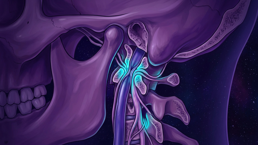
A young patient presents with a racing heart on standing, pressure behind the eyes, brain fog, and the sensation that their skull is too heavy for their neck. A cardiologist calls it POTS. A neurologist calls it migraine. A psychiatrist calls it anxiety. In a recognizable subset of these patients, all three are describing the same mechanical failure a few centimeters below the brainstem. This is the craniocervical triad: cervical instability, jugular and vagal compression, and dysautonomia braided into one structural problem.
Naming the Triad
The craniocervical triad is the co-occurrence of three findings that share a single anatomical origin at the skull base:
- Craniocervical and atlantoaxial instability — excessive movement between the skull, the atlas (C1) and the axis (C2), the joints that carry the head. When the ligaments holding these joints are lax or injured, the head shifts on the spine under normal loads.
- Jugular and vagal compression — crowding of the jugular foramen (the bony canal at the skull base through which the internal jugular vein and the vagus, glossopharyngeal, and accessory nerves exit the skull). Structures that should glide freely get pinched against bone.
- Dysautonomia — dysregulation of the autonomic nervous system, most visibly as orthostatic intolerance and postural tachycardia.
These are not three coincidental diagnoses. They are three downstream readouts of one upstream mechanical event.
The Anatomy of the Bottleneck
The skull base is the busiest few square centimeters in the body. Within a small volume the brainstem transitions to spinal cord, the vertebral arteries loop through bony rings, and the lower cranial nerves thread out alongside the great veins that drain the brain. The vagus nerve (the tenth cranial nerve and the primary parasympathetic conduit between brainstem and viscera) leaves the skull through the jugular foramen shoulder-to-shoulder with the internal jugular vein.
When the craniocervical junction is unstable, two things happen. First, the head settles or tilts into positions the anatomy never planned for, and the jugular foramen and the space behind the styloid process narrow. Second, the constant micro-motion irritates the structures passing through. A vein that is intermittently pinched cannot drain the brain efficiently; a nerve that is chronically compressed sends abnormal traffic. The result is a mechanical bottleneck where venous outflow and vagal signaling are throttled at the same choke point.
This matters because the vagus is not a bystander. Roughly 80% of its fibers are afferent (carrying information from the body toward the brain, not commands outward). Compress it and you do not just weaken an output cable — you corrupt the sensory feed the brainstem relies on to run heart rate, blood pressure, and gut motility.
The Numbers
- The diagnostic threshold for postural tachycardia is a sustained heart-rate rise of ≥ 30 bpm within 10 minutes of standing (≥ 40 bpm in adolescents), without a drop in blood pressure.
- Approximately 80% of vagal fibers are afferent — sensory — so vagal compression is largely a failure of incoming signal, not outgoing tone.
- Internal jugular veins carry the majority of cerebral venous drainage in the upright posture; when outflow is obstructed on one or both sides, intracranial pressure and symptom burden climb measurably on standing.
- Craniocervical instability clusters heavily in connective-tissue disorders: in hypermobile populations, ligamentous laxity at C1–C2 is disproportionately common, which is why the triad and heritable connective-tissue phenotypes travel together.
Why It Clusters
The common denominator is often connective tissue. The alar and transverse ligaments that stabilize the atlantoaxial joint are collagen structures. In people whose collagen is intrinsically lax — whether from a heritable disorder, whiplash injury, or chronic loading — those ligaments stretch, the joint loosens, and the skull-base geometry drifts. Once the geometry drifts, the jugular foramen narrows and the vagus is compromised. Once the vagus is compromised, the baroreflex (the moment-to-moment loop that adjusts heart rate to defend blood pressure against gravity) loses its sensory input and orthostatic control collapses. Instability is the cause; jugular-vagal compression is the mechanism; dysautonomia is the symptom.
This sequence explains the classic "unexplained" POTS presentation. The autonomic testing is abnormal, but the autonomic nerves themselves are healthy — they are simply being crushed or starved of afferent data by a structural problem the tilt-table cannot see. The pathology is orthopedic wearing an autonomic mask.
The Positional Signature
The tell is that symptoms are positional and mechanical, not random. Patients report that specific head positions — looking up, lying flat, turning to one side — predictably worsen the pressure, the tachycardia, or the fog, and that traction or a supportive collar relieves them. Purely neurochemical dysautonomia does not obey head geometry this way. When symptoms track with the position of the skull on the spine, the clinician is looking at a mechanical bottleneck, not a chemistry problem.
What This Means for Practitioners
The practical lesson is that a POTS diagnosis should not close the investigation — it should open a structural question. When orthostatic tachycardia arrives packaged with neck pain, heaviness of the head, positional headache, and connective-tissue signs, the craniocervical junction deserves attention before the case is filed as idiopathic dysautonomia or, worse, as anxiety.
- Take the mechanical history seriously. Ask whether symptoms change with head position, whether a collar helps, and whether there was a preceding neck injury. Positional dependence is a structural fingerprint.
- Screen for the connective-tissue phenotype. Hypermobility, easy bruising, and a family history point toward the laxity that lets the junction destabilize in the first place.
- Sequence the workup toward the choke point. Upright and dynamic imaging of the craniocervical junction and jugular outflow answers questions that a resting scan cannot.
- Stabilize before you medicate. When the driver is mechanical, decompressing or stabilizing the junction addresses the cause; autonomic medication only softens the readout.
The reframe is the whole point. A patient told they have three unrelated conditions in three different body systems is a patient told they are simply unlucky. A patient shown that one structural failure at the skull base drives all three is a patient with a target. The craniocervical triad turns a mystery illness into an anatomy problem — and anatomy problems have addresses.
Reference: Journal of Neurology, Neurosurgery & Psychiatry — craniocervical instability and autonomic dysfunction (2023).