Connective Tissue and the Autonomic System: How Collagen Integrity Shapes Vagal Function
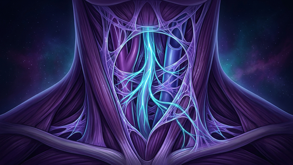
We tend to think of the autonomic nervous system as pure electricity — signals racing along nerves, reflexes firing, chemistry cascading. But every one of those nerves and vessels is a physical object living inside a physical scaffold. Blood vessels have walls that must hold their shape against pressure. The vagus nerve runs through a sleeve of tissue that must cushion and anchor it. That scaffold is connective tissue, and its master material is collagen. When the collagen is faulty — as it is in the hypermobility spectrum and the Ehlers-Danlos syndromes — the autonomic system does not fail because the wiring is broken. It fails because the building the wiring lives in is structurally unsound. This is dysautonomia as an architecture problem.
Collagen is the scaffold, not the decoration
Collagen is the most abundant protein in the body, making up roughly a third of total protein mass, and it is the primary structural fiber of connective tissue (the material that holds organs, vessels, and nerves in their proper shape and place). It is the rebar in the concrete: it gives blood-vessel walls their resilience, ligaments their restraint, and the sheaths around nerves their protective firmness.
In the heritable connective tissue disorders — the Ehlers-Danlos syndromes chief among them — the genetic instructions for building or processing collagen are altered. The resulting fibers are laid down disorganized, over-stretchy, or fragile. The visible consequence is joint hypermobility (joints that move well beyond their normal range) and skin that stretches too far. But collagen is everywhere the visible tissue is not, and the invisible consequences — inside the vessels and around the nerves — are where the autonomic trouble begins.
Distensible veins and the physics of blood pooling
Start with the vasculature, because it is the clearest mechanical link. Vein walls owe their tone and recoil substantially to collagen and elastin. When those fibers are lax, the veins become abnormally distensible (able to stretch and balloon under pressure that a normal vein would resist).
Now apply gravity. The moment a person with over-compliant veins stands, blood does what physics dictates — it falls into the roomy, low-resistance vessels of the legs and abdomen and pools there instead of returning promptly to the heart. Studies have estimated that on standing, on the order of 500 to 700 milliliters of blood can shift into the lower body; in a compliant venous system that pooling is exaggerated. Less blood returns to the heart, so stroke volume drops, and the autonomic system must compensate — it fires a hard sympathetic burst to slam the heart rate up and defend blood pressure to the brain. That compensatory tachycardia is precisely the definition of postural orthostatic tachycardia syndrome (POTS): a sustained heart-rate rise of at least 30 beats per minute on standing without a matching drop in blood pressure. In a large fraction of hypermobile patients, POTS is not a separate disease that happens to co-occur. It is the direct hemodynamic consequence of collagen that cannot hold the veins to shape.
The carotid sheath: where the vagus lives
Now the nerve itself. In the neck, the vagus nerve does not travel alone. It runs inside the carotid sheath (a firm connective-tissue tube that bundles the carotid artery, the internal jugular vein, and the vagus nerve together and fixes them in position). That sheath is connective tissue. Its job is to hold the nerve in a stable, protected channel and to buffer it against the constant motion of the neck, the pulsation of the artery beside it, and the pressure of the tissues around it.
When the sheath and the surrounding fascia are built from lax collagen, the nerve loses its stable housing. Two things follow. First, the vagus becomes mechanically vulnerable — more exposed to traction as the neck moves, more susceptible to compression from a shifting cervical structure, less buffered against the mechanical insults a firm sheath would absorb. Mechanotransduction (the conversion of mechanical force into a biological or electrical signal) means a nerve under abnormal stretch or pressure does not stay silent; it misfires, and its baseline signaling degrades. Second, lax ligaments in the neck permit craniocervical and cervical instability (excess movement between the skull and upper vertebrae), which places the brainstem, the jugular outflow, and the vagal roots under mechanical strain that a stable neck would never impose. The nerve is not diseased. It is being manhandled by the structure that was supposed to protect it.
One tissue defect, a cluster of autonomic failures
Trace a single faulty protein outward and a whole autonomic pattern falls into place — a connective-tissue-to-dysautonomia axis in which the tissue defect is upstream of everything:
- Orthostatic intolerance and POTS — from distensible veins and exaggerated blood pooling on standing.
- Blunted baroreflex buffering — the baroreflex (the pressure-sensing loop that stabilizes blood pressure) relies on stiff, responsive arterial walls; over-compliant vessels blur the very signal it depends on.
- Mechanical vagal vulnerability — from an unstable carotid sheath and cervical instability stressing the nerve and its roots.
- Poor venous return and low filling pressure — leaving the whole system chronically volume-challenged and sympathetically overdriven.
The numbers underline how tightly these travel together. Dysautonomia, and POTS in particular, is strikingly over-represented in hypermobile and Ehlers-Danlos populations — surveys commonly report that a large majority of hypermobile-EDS patients meet criteria for orthostatic intolerance — and POTS itself skews heavily female, roughly 4 to 5 patients in 5 being women, mirroring the demographics of the hypermobility spectrum. When two conditions share both a mechanism and a demographic this closely, coincidence is the least likely explanation.
Why the workup keeps coming back normal
This is a structural and mechanical problem, and standard autonomic and cardiac testing is built to catch electrical and endocrine disease. An echocardiogram reads normal because the heart muscle is fine. Bloodwork reads normal because there is no inflammatory or hormonal lesion to find. The defect is in the compliance of the vessel wall and the integrity of the nerve's housing — properties that a resting ECG and a chemistry panel simply do not measure. The patient is told they are healthy while living inside a scaffold that cannot do its mechanical job. Recognizing the connective-tissue substrate is often what finally explains a decade of "normal" tests.
What this means for practitioners
If the substrate is mechanical, the management is substantially mechanical — you are compensating for a scaffold that cannot hold shape on its own.
- Screen the connective tissue. In any dysautonomia or POTS presentation, assess joint hypermobility with a Beighton score and ask about skin, easy bruising, and family history. The tissue phenotype reframes the whole picture.
- Give the veins external support. Because the vein walls cannot recoil on their own, compression garments and abdominal binding physically restrain the pooling, and aggressive fluid and sodium loading raises the filling volume the lax system loses. These are mechanical fixes for a mechanical fault.
- Protect and stabilize the neck. Targeted strengthening of the deep cervical stabilizers substitutes muscular control for the ligamentous restraint the collagen cannot provide, reducing the mechanical stress on the vagus and its roots.
- Support the collagen substrate. Adequate protein and the cofactors collagen synthesis requires — vitamin C prominent among them — give the body the raw materials to lay down the best fiber its genetics allow.
- Rebuild vagal tone deliberately. Slow-breathing work and HRV tracking raise parasympathetic tone to counterbalance the chronic sympathetic overdrive that lax vessels and an unstable neck impose.
Reference: American Journal of Medical Genetics — hypermobility, connective tissue, and dysautonomia (2021).