Cervicovagopathy: How an Unstable Neck Compresses the Vagus and Drives Dysautonomia
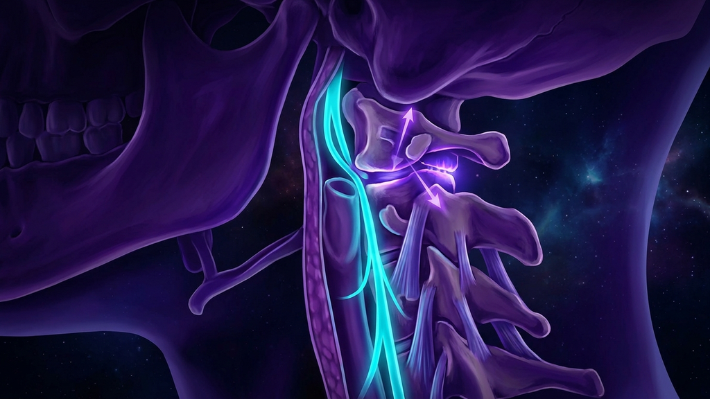
A patient stands and their heart rate leaps 40 beats. They turn their head and feel faint. They spend years being told it is panic. But the vagus nerve does not run through open space — it descends through the neck millimeters from the top two vertebrae, and if those vertebrae move too far, the nerve is compressed on every motion. When neck mechanics drive vagal dysfunction, the condition has a name: cervicovagopathy. It is the single most important structural reframe in dysautonomia, and it is not the same thing as a tight neck.
What Cervicovagopathy Actually Is
Cervicovagopathy (vagus nerve compression or irritation within the neck) describes a mechanical problem being misread as a neurological or psychiatric one. The vagus (cranial nerve X) travels the length of the neck inside a connective-tissue tube alongside the carotid artery and jugular vein, passing directly beside the upper cervical vertebrae. When the structures around it shift — through instability, rotation, or inflammation — the nerve is stretched, kinked, or pressed against bone. The result is not "a knot in the muscle." It is disordered signaling in the body’s master autonomic cable.
This is the critical distinction from generic neck tension. Muscular tightness is a soft-tissue complaint. Cervicovagopathy is a neuromechanical one: the symptoms are autonomic — heart rate, blood pressure, digestion, air hunger — because the compressed structure is an autonomic nerve, not a muscle.
The Instability Underneath: C1-C2
The most common mechanical driver is instability at the top of the spine. Two overlapping terms matter:
- Craniocervical instability (CCI) — excessive movement between the skull and the first vertebra, where the head is inadequately anchored to the neck.
- Atlantoaxial instability (AAI) — excessive movement between C1 (the atlas) and C2 (the axis), the joint responsible for roughly 50% of the neck’s total rotation.
Because the C1-C2 segment handles about half of all head rotation, it is uniquely exposed. Every time the head turns, structures at this level move — and if the stabilizing ligaments are lax, they move too far. Radiographic thresholds make this concrete: an atlantodental interval exceeding roughly 3 mm in adults, or a few millimeters of pathological translation, marks instability. Millimeters are the difference between a nerve that glides and a nerve that is pinched.
Why an Unstable Neck Attacks the Vagus
When the atlas slides or rotates beyond its normal range, several things happen at once:
Direct mechanical load
The vagus and the internal jugular vein sit just anterolateral to the C1 transverse process. A forward-slipped or rotated atlas presses the sheath and its contents against unyielding bone, particularly on head rotation — the reason symptoms are so reliably positional.
Venous outflow obstruction
The same instability that compresses the nerve compresses the jugular vein beside it, throttling cerebral venous drainage. The resulting congestion feeds brain fog, pressure headache, and the wired-and-tired state, and it raises the mechanical pressure on the adjacent vagus.
Baroreflex disruption
Distorted vagal and glossopharyngeal signaling degrades the baroreflex (the loop that adjusts heart rate and vessel tone to keep blood pressure stable when you change posture). A degraded baroreflex is exactly what produces orthostatic tachycardia — the heart-rate rise of 30 bpm or more on standing that defines POTS.
From "Anxiety" to Structure
This is the pivot that changes patients’ lives. The classic dysautonomia presentation — racing heart, lightheadedness, air hunger, brain fog, tremor, GI slowing — is the exact symptom set of a compressed vagus. When those symptoms track with head position, worsen with neck loading, and appear in someone with a hypermobile or connective-tissue background, an "anxiety disorder" is very likely a mislabeled cervicovagopathy. The nervous system is not overreacting to nothing; it is reacting to a wire under mechanical stress.
The Connective-Tissue Link
Instability is not random. In heritable connective-tissue disorders — most prominently hypermobile Ehlers-Danlos syndrome (hEDS) (a collagen disorder that weakens ligaments and vessel walls) — the ligaments that should anchor C1 and C2 are lax by biology. This is why cervicovagopathy clusters with hypermobility and POTS: roughly 80% of hEDS patients have autonomic dysfunction, and the shared thread is connective tissue that will not hold the neck, the vessels, or the nerve sheath in place. The instability and the dysautonomia are two faces of one collagen problem.
Why Upright, Dynamic Imaging Matters
Here is the diagnostic trap. Standard MRI is performed supine and still — the one position in which an unstable neck looks normal, because gravity and rotation are removed. A ligament that fails only under load is invisible to an unloaded scan. This is why so many patients accumulate "normal" imaging and a psychiatric label.
The mechanism reveals itself only under provocation: upright or dynamic imaging (flexion-extension and rotational views taken with the neck loaded or moving) can expose translation that supine films miss entirely. If the complaint is positional, the imaging must be positional too. Reproducing symptoms with the structure under real-world load is the whole point.
What This Means for Practitioners
- Ask about position. Symptoms that change with head turning, looking up, or lying on one side point toward the neck, not the psyche.
- Screen the connective tissue. A hypermobility assessment reframes the whole picture and predicts who is at risk for instability.
- Image under load. A supine "normal" MRI does not exclude craniocervical or atlantoaxial instability; dynamic views are required to see it.
- Stabilize before you sedate. Interventions that reduce upper-cervical load and support the deep neck stabilizers target the driver; anxiolytics target the symptom.
Cervicovagopathy is the definitive structural explanation for a huge share of "unexplained" dysautonomia. The vagus is being compressed by a neck that moves a few millimeters too far. Name it, load the imaging, and the anxiety diagnosis dissolves into anatomy.
Reference: Frontiers in Neurology, craniocervical instability and dysautonomia (2023).