Ultrasound at the Subdiaphragmatic Branches: A Non-Invasive Route into the Inflammatory Reflex
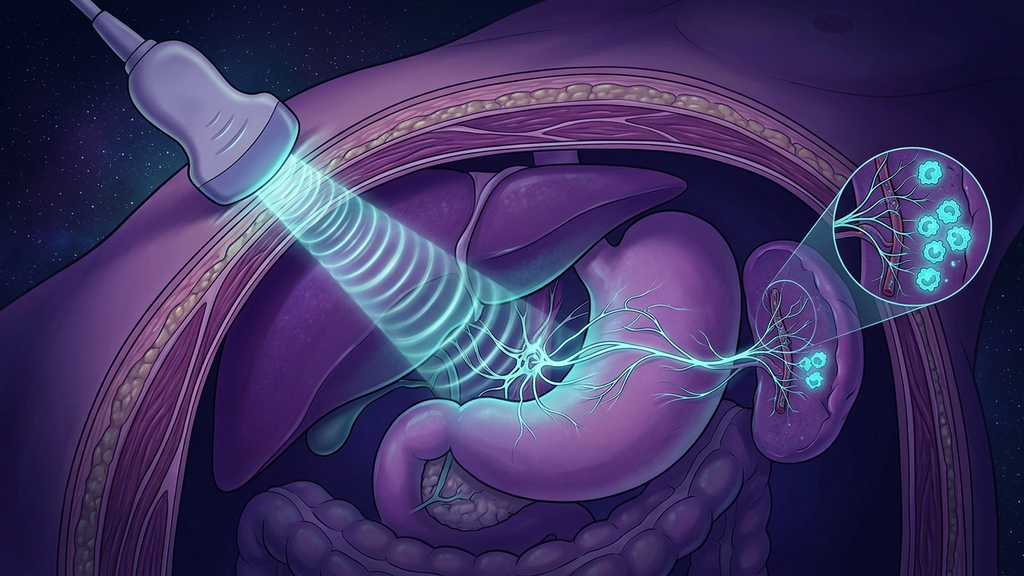
The inflammatory reflex has a well-mapped off-switch, but flipping it has usually meant surgery: an electrode cuffed around the vagus in the neck, wired to an implanted pulse generator. Effective, but invasive. Over the last decade a different idea has been tested in animals: what if you could reach the same circuit from the outside, using focused sound? The target is not the neck at all. It is the vagal branches below the diaphragm, and the organ they lead to. This is an emerging, largely preclinical story, and it deserves to be told with its caveats intact, but the mechanism is clean enough to take seriously.
Why Aim Below the Diaphragm
The neck is the classic access point, but it is crowded and the whole cervical vagus fires together, producing off-target effects on heart rate and the airway. The subdiaphragmatic vagus (the vagal branches running below the diaphragm to the abdominal organs) offers a more selective target, because here the fibers have largely sorted toward specific destinations, including the pathway that governs the spleen. Reaching this level means engaging the anti-inflammatory arm with less collateral effect on the cardiac branches above.
And the spleen itself is reachable. The splenic neurovascular bundle (the nerve fibers that travel with the splenic artery into the organ) sits in the upper abdomen, accessible to an acoustic beam aimed through the body wall in a way the deep neck fibers are not.
How Sound Becomes a Neural Signal
The tool is focused ultrasound (high-frequency sound waves converged to a small target point inside the body without any incision). The same physics that lets ultrasound image a fetus lets a focused beam deposit mechanical energy at a precise depth. When that energy is delivered to nerve-rich tissue, it can mechanically excite the fibers there, a process sometimes called sonication of the target.
The proposed chain is specific and testable:
- Focused ultrasound is aimed at the splenic or subdiaphragmatic vagal fibers.
- The mechanical stimulus activates the cholinergic anti-inflammatory pathway (the acetylcholine-mediated circuit that ends with macrophages being told to stop releasing cytokines).
- Splenic macrophages downshift, and the measured output, serum TNF-alpha (tumor necrosis factor, a lead inflammatory cytokine), falls.
The elegance is that modality, mechanism, and outcome form a single legible line: sound in, cytokine down.
What the Preclinical Literature Actually Shows
The evidence base here is animal work and early translational studies, and it should be described as such.
- In rodent models of inflammatory challenge, ultrasound stimulation aimed at the spleen has reduced circulating TNF and related cytokines by roughly 50-70% compared with sham stimulation.
- The effect has been shown to depend on the pathway: severing the splenic nerve or blocking the relevant receptors abolishes it, arguing the ultrasound is working through the neural circuit rather than by generic tissue heating.
- Related work has reported that splenic ultrasound can blunt other pathway-dependent responses, such as inflammation-driven changes in glucose handling, in animal models.
- Most protocols use brief exposures, on the order of minutes per session, at intensities intended to stimulate rather than ablate tissue.
Why the Subdiaphragmatic Target Is Clever
The choice of target is the most interesting engineering decision in this whole line of work. Stimulating the cervical vagus in the neck is like flipping a master switch: you get the anti-inflammatory effect, but you also tug on the fibers that set heart rate and bronchial tone, which is why implanted neck stimulators are dosed carefully around cardiac side effects. Below the diaphragm, the fiber populations have largely diverged toward their organ destinations, so a stimulus placed there can, in principle, favor the splenic anti-inflammatory arm while sparing the cardiac branches that sit safely upstream in the chest and neck. Selectivity, not just non-invasiveness, is the real prize.
There is a second advantage. Because the spleen concentrates such a large share of the body's inflammatory cells in one place, a beam that reaches the splenic bundle acts on a natural bottleneck: a small, well-chosen target with a disproportionately large downstream effect on systemic cytokine output.
The Caveats, Stated Plainly
Enthusiasm has to be bounded by honesty. Nearly all of the mechanistic proof is in animal models; human data are early and far smaller. Targeting is genuinely hard: the spleen and vagal branches move with breathing and vary in position between people, so aiming a focused beam reliably at a small deep structure is a real engineering problem. Dose-response, durability, and safety windows in humans are not yet settled. And "ultrasound reduced a cytokine in a mouse" is not the same claim as "ultrasound treats an inflammatory disease in a person." The correct posture is cautious interest, not conviction.
Why It Still Matters
Even held to that standard, the approach is important for one reason: it decouples access to the inflammatory reflex from surgery. If a focused beam can engage the spleen-brain axis transiently and non-invasively, then the body's own anti-inflammatory brake becomes something you can reach without implanting hardware. That is a genuinely different therapeutic geometry, and it is why the subdiaphragmatic and splenic ultrasound literature is worth watching closely.
The Takeaway for Practitioners
The concept to hold is the chain, not the hype: a non-invasive mechanical stimulus, aimed at a specific neuroanatomical target below the diaphragm, engaging a defined immune circuit, with a measurable cytokine readout. That framing keeps the modality tethered to mechanism and outcome, and it keeps expectations tethered to the fact that, for now, most of the strongest evidence still comes from the lab bench and the animal model, not the clinic.
Reference: preclinical splenic and peripheral focused-ultrasound neuromodulation studies, Nature Communications and related journals (2019-2023).