Exercise as Mitochondrial Medicine: How Movement Literally Builds New Powerhouses
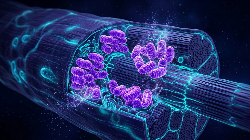
Of all the interventions we have for cellular energy production, only one has been shown to literally grow new mitochondria. Not supplements, not light therapy, not pharmacology. Exercise. The mechanism is well characterized, the dose-response is mappable, and yet the prescription is one of the most poorly delivered tools in medicine — because the same exercise that builds mitochondria in a healthy patient destroys them in a chronically ill one.
The Molecular Switch: PGC-1α
The master regulator of mitochondrial biogenesis is a transcriptional coactivator called PGC-1α (peroxisome proliferator-activated receptor gamma coactivator 1-alpha). When skeletal muscle contracts, intracellular calcium rises, AMP/ATP ratio rises, and ROS signaling rises — all three converge on PGC-1α activation. PGC-1α then turns on the transcription of mitochondrial proteins encoded in both nuclear and mitochondrial DNA. The net effect: more mitochondria, larger mitochondria, and improved mitochondrial respiratory capacity.
The Two Stimuli That Drive Biogenesis
Zone-2 endurance training
Sustained aerobic effort at 60–70% of maximum heart rate — the "you could hold a conversation but you would not want to" intensity. Zone-2 sessions of 60–90 minutes, performed 3–4 times per week, have been shown to increase mitochondrial content by 40–50% over 12 weeks. The mechanism is sustained calcium and AMPK signaling driving PGC-1α activation. Zone 2 also preferentially trains slow-twitch fibers, which are mitochondrion-rich by design.
High-intensity interval training (HIIT)
Repeated bouts of near-maximal effort (e.g., 4×4-minute intervals at 90% HRmax, with 3-minute recoveries). HIIT increases nuclear abundance of PGC-1α acutely and has been shown to increase mitochondrial respiratory capacity by 49% over six weeks in previously untrained adults. The 2025 evidence is clear that HIIT and Zone 2 produce comparable PGC-1α responses, though HIIT does so in a fraction of the time.
"HIIT has superior effectiveness in mitochondrial biogenesis and volume density per unit of training time, making it a viable alternative to moderate-intensity continuous training — particularly for time-constrained patients with intact baseline mitochondrial capacity."
What the Combined Approach Looks Like
The most robust mitochondrial training stack pairs Zone 2 for volume with HIIT for intensity:
- 2–3 Zone-2 sessions per week, 45–90 minutes each.
- 1–2 HIIT sessions per week, 20–30 minutes each.
- 2–3 resistance training sessions per week — important for muscle quality and mitochondrial density per fiber.
- One full rest day weekly. Adaptation happens in recovery, not in training.
The Critical Caveat: Mitochondrial Capacity First
Here is where most exercise prescriptions go wrong. The biogenesis response requires intact baseline mitochondrial function. In patients with ME/CFS, long COVID, advanced CIRS, or severe burnout, the mitochondria cannot meet the energy demand of conventional training. The result is post-exertional malaise — a multi-day crash in which symptoms worsen and recovery markers (HRV, sleep, cognitive function) deteriorate.
In these patients, the rule is different:
- Start with recumbent or seated activity at very low intensity (Zone 1).
- Use heart rate to cap effort, not perceived exertion (perception is unreliable in this group).
- Monitor next-day response. If recovery markers worsen, dial back.
- Build duration before intensity.
- Only when baseline mitochondrial capacity has been restored should Zone 2 or interval work enter the plan.
Beyond the Muscle: Brain Mitochondria
Exercise-induced mitochondrial biogenesis is not limited to skeletal muscle. Aerobic exercise increases hippocampal mitochondrial biogenesis, doubles the rate of adult neurogenesis in animal models, and is associated with improved cognitive performance in humans. The cellular mechanism is the same — PGC-1α activation — but the clinical implication is profound: training for muscle mitochondria simultaneously trains brain mitochondria.
Cofactors That Amplify the Response
Several nutritional supports amplify training-induced biogenesis:
- Creatine monohydrate (5 g/day) — supports phosphocreatine buffering, particularly relevant for HIIT.
- CoQ10 (ubiquinol) — preserves electron transport chain efficiency.
- Magnesium glycinate — cofactor for ATP synthase.
- NAD+ precursors (NR or NMN) — emerging evidence for support of sirtuin signaling, which interacts with PGC-1α.
- Polyphenols (resveratrol, quercetin, urolithin A) — activate mitochondrial biogenesis and mitophagy.
The New-Medicine Adjuncts
Photobiomodulation (red and near-infrared light) directly stimulates cytochrome c oxidase in the electron transport chain. PEMF therapy has shown ATP elevations in cell-culture and animal models. Neither replaces exercise — they support it, particularly during recovery phases or in patients whose tolerance for conventional training is limited.
Clinical takeaway: Movement is the only intervention that grows new mitochondria. Dose it correctly — Zone 2 for volume, HIIT for intensity, resistance training for muscle quality. In healthy patients, push the prescription. In ME/CFS, long COVID, or severe burnout, build mitochondrial capacity at the recovery edge first, or you will spend months in post-exertional flares.
References
- Granata C et al. "High-intensity training induces non-stoichiometric changes in the mitochondrial proteome of human skeletal muscle." Nature Communications, 2021;12:7056.
- Little JP et al. "An acute bout of high-intensity interval training increases the nuclear abundance of PGC-1α and activates mitochondrial biogenesis." American Journal of Physiology, 2011;300(6):R1303-R1310.
- Memme JM et al. "Exercise and mitochondrial health." Journal of Physiology, 2021;599(3):803-817.
- Bishop DJ et al. "High-intensity exercise and mitochondrial biogenesis: current controversies and future research directions." Physiology, 2019;34(1):56-70.
- Drake JC et al. "Molecular mechanisms for mitochondrial adaptation to exercise training in skeletal muscle." FASEB Journal, 2016;30(1):13-22.