Electromagnetic Therapy and the Cellular Antenna: How Fields Modulate Living Tissue
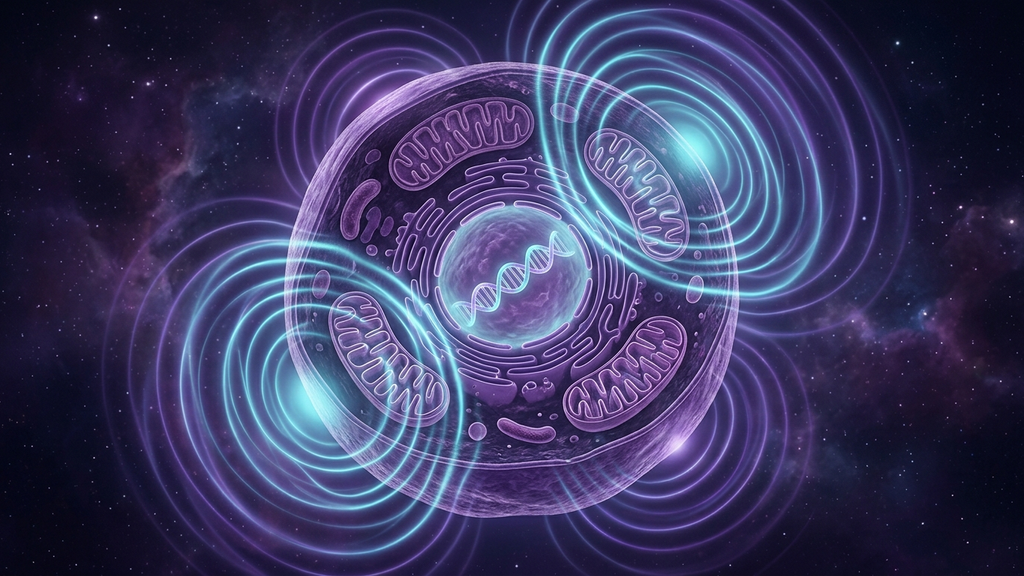
For most of the twentieth century, the dominant model in medicine was chemical. Disease was a problem of molecules in the wrong amounts and the wrong places. Treatment was the science of correcting those molecular imbalances. This model has been productive — pharmacology has saved lives at an unprecedented scale — but it is incomplete. The cell is not only a chemical system. It is also an electrical and electromagnetic one. And the fields that surround it are not background noise — they are part of the signaling environment that determines what the cell actually does.
That recognition is the foundation of electromagnetic therapy, an umbrella term that covers a growing family of clinical tools: pulsed electromagnetic field (PEMF) therapy, transcranial magnetic stimulation, focused ultrasound, photobiomodulation, scalar field devices, and bioelectronic medicine more broadly.
Why Cells Respond to Electromagnetic Fields
Every cell maintains a transmembrane potential — typically –40 to –90 millivolts across the plasma membrane. Mitochondria maintain their own membrane potential, which is essential for ATP production. DNA itself produces electromagnetic signaling, and microtubules inside neurons appear to oscillate at frequencies ranging from kilohertz to terahertz. The cellular environment is not just chemically active — it is a hum of electromagnetic activity.
Externally applied electromagnetic fields, in the right frequency ranges and intensities, can:
- Modulate ion channel kinetics, particularly voltage-gated calcium channels
- Influence mitochondrial membrane potential and ATP production
- Affect gene expression through electromagnetic transduction pathways
- Modulate cytokine release and immune signaling
- Influence neural firing rates non-invasively
These are not exotic claims. Each of these mechanisms has been demonstrated in vitro and replicated across labs. The clinical question is no longer whether electromagnetic fields affect cells — they clearly do — but which fields, in what doses, for which conditions.
PEMF: The Most Mature of the Tools
Pulsed electromagnetic field therapy has FDA clearance for fracture non-union (since 1979) and for several other applications including post-surgical edema and depression-adjunct treatment. The mechanism appears to involve:
- Increased nitric oxide signaling, supporting vascular health
- Modulation of mitochondrial function and ATP production
- Influence on osteoblast and fibroblast activity, supporting tissue repair
- Reduced inflammatory cytokine production
Clinical signal is strongest in orthopedic applications, but reasonable evidence exists for use in chronic pain, depression-adjunct, post-concussion recovery, and fatigue states. The therapeutic windows are narrow — frequency, intensity, and exposure duration all matter — which is part of why PEMF results in the literature are sometimes inconsistent.
Transcranial Magnetic Stimulation (TMS)
TMS delivers focused magnetic pulses through the skull to induce neural firing in targeted cortical regions. It has FDA clearance for major depressive disorder, OCD, and several other neuropsychiatric conditions. The mechanism is electromagnetic induction — a rapidly changing magnetic field produces local electrical currents that depolarize neurons. With sustained treatment, neuroplastic changes occur, and the underlying circuit dysfunction begins to remodel.
Focused Ultrasound — Mechanical Rather than Electromagnetic
While not strictly electromagnetic, focused ultrasound is increasingly grouped with these technologies because it shares the same underlying logic — physical-field modulation of cellular function. Low-intensity focused ultrasound can modulate neural firing through mechanotransductive effects on ion channels, and it can do so with much greater spatial precision than TMS. The therapeutic applications under investigation range from anxiety and depression to non-invasive vagus nerve stimulation to neuroinflammation modulation.
Photobiomodulation
Red and near-infrared light at specific wavelengths is absorbed by cytochrome c oxidase in the mitochondrial inner membrane, stimulating ATP production and modulating reactive oxygen species signaling. Clinical applications include wound healing, musculoskeletal injury, traumatic brain injury recovery, depression-adjunct, and skin health. The non-invasive nature and excellent safety profile make this one of the most accessible of the new tools.
Scalar and Quantum-Field Devices
This is the most controversial category. Devices marketed under terms like "scalar" or "biofield" claim to deliver non-Hertzian electromagnetic signals that influence cellular function. The theoretical mechanisms — drawn variously from Tesla, biofield physics, and quantum biology — are not well-validated. Some products in this category appear to deliver real low-frequency magnetic fields that may have measurable biological effects; others appear to do nothing. The field is undisciplined. A serious clinician evaluating a scalar device should ask: what frequencies and intensities are actually being delivered, and is there third-party measurement to support the marketing claims?
Bioelectronic Medicine: The Implantable Frontier
At the most invasive end of the spectrum sits bioelectronic medicine — implanted devices that deliver targeted electrical stimulation to specific nerves. Vagus nerve stimulators (Cyberonics/LivaNova) for epilepsy and depression. SetPoint Medical's implant for rheumatoid arthritis, which delivers vagal stimulation to suppress inflammation. Sacral nerve stimulators for bladder dysfunction. This is the chemical-free pharmacology of the future — electrical signals delivered with the precision of a drug.
The Combined Approach
In clinical practice, these tools are increasingly being layered rather than chosen between. A typical protocol for a long COVID or post-concussion patient might include daily photobiomodulation to the head and neck, scheduled PEMF sessions over the chest and pelvis, focused ultrasound for cervical vagal stimulation, and standard mitochondrial and vagal support strategies running in parallel. The reasoning is mechanistic: each tool addresses a slightly different part of the cellular-energy and signaling cascade, and the combination is more effective than any single intervention alone.
A Word on Quality Control
The electromagnetic-therapy market is not well-regulated, and product quality varies widely. The questions worth asking before recommending or purchasing any device:
- What frequencies and intensities does it actually deliver, and has this been measured by an independent lab?
- Is there peer-reviewed clinical evidence for the specific application, or only marketing claims?
- Is the manufacturer transparent about the underlying mechanism, or does the marketing rely on quantum jargon without specific physical claims?
- Is the device FDA-cleared (where applicable) for any indication?
A serious electromagnetic therapy device should pass all four. Many of the most heavily marketed devices in this space pass only one or two.
The big picture: Cells are electromagnetic structures, and they respond to applied electromagnetic fields. That is no longer in serious scientific dispute. The clinical question is precision — which fields, at what doses, for which conditions. The mature tools (PEMF, TMS, photobiomodulation, focused ultrasound) have earned a place in the toolkit. The less mature tools are worth following but not yet ready to anchor a treatment plan.
References
- Markov MS. "Pulsed electromagnetic field therapy: history, state of the art and future." The Environmentalist, 2007;27(4):465-475.
- Lefaucheur JP et al. "Evidence-based guidelines on the therapeutic use of repetitive transcranial magnetic stimulation (rTMS)." Clinical Neurophysiology, 2020;131(2):474-528.
- Hamblin MR. "Mechanisms and applications of the anti-inflammatory effects of photobiomodulation." AIMS Biophysics, 2017;4(3):337-361.
- Koopman FA et al. "Vagus nerve stimulation inhibits cytokine production and attenuates disease severity in rheumatoid arthritis." PNAS, 2016;113(29):8284-8289.
- Tyler WJ. "The mechanobiology of brain function." Nature Reviews Neuroscience, 2012;13(12):867-878.