Brain Fog Decoded: The Mitochondrial and Vagal Roots of Foggy Thinking
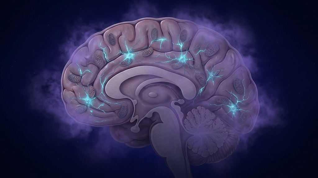
"Brain fog" is one of the most common — and most dismissed — complaints I see in clinic. Patients describe it as thinking through molasses, losing words mid-sentence, reading the same paragraph three times. They are usually told it is stress, age, or anxiety. The mechanistic picture is more specific: brain fog is a measurable failure of cerebral energy production, amplified by neuroinflammation and autonomic imbalance.
The Bioenergetics of Cognition
The brain is 2% of body weight and consumes roughly 20% of total energy at rest. Cognition is metabolically expensive — sustained attention alone increases regional glucose uptake by 20–30%. When mitochondria in cortical neurons cannot meet that demand, executive function, working memory, and processing speed are the first to degrade. The patient experiences this as fog.
The Three Mechanisms Behind Most Cases
In clinic, brain fog almost always traces back to one or more of these pathways:
- Mitochondrial dysfunction. Reduced complex I and IV activity, oxidative stress, and impaired ATP output have been documented in long COVID, ME/CFS, depression, and post-infectious syndromes — exactly the populations who present with persistent fog.
- Neuroinflammation. Activated microglia release IL-6 and TNF-α, which directly impair synaptic transmission and slow neural processing. 2025 reviews of long COVID brain fog identify microglial priming and NLRP3 inflammasome activation as central drivers.
- Autonomic dysregulation. A nervous system stuck in sympathetic dominance shunts blood flow away from the prefrontal cortex. The patient is "alert" but cognitively impaired — the classic anxious-but-foggy presentation.
Why the Standard Workup Misses It
A normal CBC, TSH, B12, and brain MRI tell you very little. Brain fog rarely shows up on coarse-grained tests. Markers worth tracking instead: hs-CRP, ferritin (often low or paradoxically high), homocysteine, fasting insulin, morning cortisol curve, and HRV. None of these are pathognomonic — but together they outline the bioenergetic and inflammatory picture.
The Treatment Stack That Actually Works
I sequence interventions in three layers.
Layer 1: Reduce neural inflammatory load
- Omega-3 fatty acids (EPA-dominant, 2–3 g/day) — reduces microglial activation.
- Sleep prioritization — glymphatic clearance occurs during deep sleep; impaired sleep is impaired brain detox.
- Identify and remove inflammatory inputs: chronic infections (EBV reactivation, Lyme, mold), gut barrier dysfunction, ultra-processed food.
Layer 2: Restore mitochondrial output
- CoQ10 (ubiquinol form, 100–200 mg/day) — supports the electron transport chain directly.
- Magnesium glycinate — cofactor for ATP synthase.
- B-complex (especially B1, B2, B3, methylated B12) — multiple cofactor roles in the TCA cycle and ETC.
- Creatine monohydrate (5 g/day) — emerging evidence for cognitive benefit, particularly in low-energy states.
Layer 3: Restore autonomic balance
- Slow nasal breathing at six breaths per minute — measurably increases HRV within a single session.
- Transcutaneous auricular vagus nerve stimulation (taVNS) — shifts autonomic balance toward parasympathetic dominance and reduces cortisol release.
- Morning sunlight and circadian alignment — recalibrates the central pacemaker that governs both autonomic and mitochondrial cycles.
Where Ultrasound Fits
Low-intensity transcranial focused ultrasound is being studied as a non-invasive way to modulate specific deep brain networks — including the default mode network, often hyperactive in foggy patients. It is an early-stage but mechanistically rational tool, and it converges with the broader bioelectronic medicine thesis: cognition is a network phenomenon, and networks respond to physical signals, not only chemical ones.
Clinical takeaway: Brain fog is rarely psychological. It is the conscious experience of cortical neurons that cannot make enough ATP, surrounded by activated microglia, under an autonomic nervous system stuck in alert mode. Treat all three, in order, and the fog lifts.
References
- Wood E et al. "Long Covid brain fog: a neuroinflammation phenomenon?" Oxford Open Immunology, 2023;4(1):iqac007.
- Lyra E Silva NM et al. "Pathogenesis of long-term neuropsychiatric COVID-19: role of microglia, mitochondria, and persistent neuroinflammation." Translational Psychiatry, 2021;11:355.
- Cuberos-Paredes E et al. "Transcutaneous auricular vagus nerve stimulation inhibits mental stress-induced cortisol release." Physiological Reports, 2025;13(9):e70251.
- Frangos E et al. "Non-invasive access to the vagus nerve central projections via auricular stimulation." Brain Stimulation, 2015;8(3):624-636.
- Picard M, McEwen BS. "Psychological stress and mitochondria: a conceptual framework." Psychosomatic Medicine, 2018;80(2):126-140.