Acid Reflux and GERD: The Vagal–Mitochondrial Roots of a Misunderstood Symptom
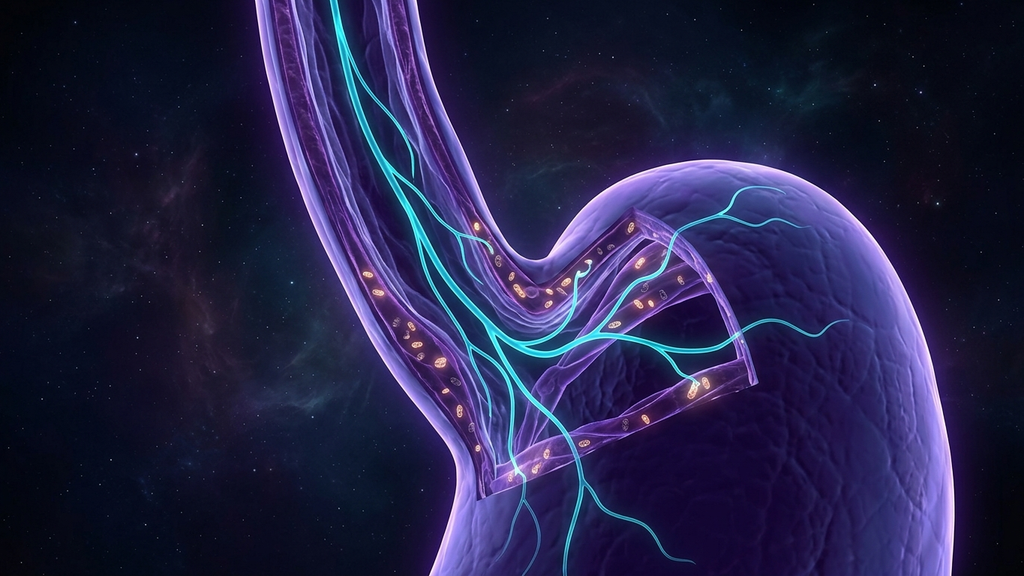
Roughly one in three adults reports chronic acid reflux. The dominant treatment — proton pump inhibitors — works on the assumption that the underlying problem is excess gastric acid. But the longer I work with patients who do not respond to acid suppression, the clearer it becomes: in the majority of cases, reflux is not an acid problem. It is a motility problem. And motility is downstream of two things you can actually treat — vagal tone and mitochondrial energy in upper-GI smooth muscle.
The Sphincter Is a Muscle, and Muscles Need Energy
The lower esophageal sphincter (LES) is a ring of smooth muscle that has to maintain tonic contraction throughout the day to keep gastric contents below it. Tonic contraction is metabolically expensive. The smooth-muscle cells of the LES are densely packed with mitochondria, and when those mitochondria are compromised — by chronic stress, micronutrient depletion, mold exposure, post-viral injury, or simply long-standing autonomic dysregulation — the sphincter loses its baseline tone. Pressure that would normally be contained spills upward into the esophagus. The patient describes burning, regurgitation, hoarseness, or that distinct sensation of "something stuck in my throat."
This is why so many patients on PPIs continue to have symptoms. The acid is being suppressed. The mechanical failure is not.
The Vagus Is the Conductor
The vagus nerve provides parasympathetic innervation to the entire upper GI tract — esophagus, stomach, LES, pyloric sphincter, and the small intestine. It coordinates the peristaltic wave that clears the esophagus after a swallow. It modulates LES pressure in coordination with gastric distention. It tells the stomach when to empty.
When vagal tone collapses — and it does in chronic stress, long COVID, POTS, ME/CFS, post-surgical recovery, and after years of sympathetic dominance — every one of these functions degrades. Peristalsis becomes uncoordinated. The LES relaxes when it should contract. Gastric emptying slows. Reflux is the symptom; vagal failure is the engine.
How to Recognize a Vagal-Motility Reflux
This phenotype has a distinct clinical signature:
- Reflux that is worse when stressed or anxious, not after meals alone.
- Bloating, early satiety, and a sense that food "sits" in the stomach.
- Low resting heart rate variability.
- Concurrent symptoms in other vagally-innervated organs: irregular bowel habits, lightheadedness on standing, cold hands and feet.
- Incomplete response to PPIs, or rebound when they are stopped.
The Bioenergetic-Vagal Protocol
I treat this phenotype in three parallel layers.
1. Restore Vagal Drive
Slow nasal breathing at approximately six breaths per minute, with extended exhales, is the cheapest and most reliable vagal intervention available. Cold-water face immersion before meals can pre-activate the dive reflex. Auricular vagus nerve stimulation — manual, transcutaneous, or via low-intensity ultrasound — is increasingly part of our clinical toolkit. Gargling, humming, and singing also activate vagal motor branches that share innervation with the upper esophagus, and these are not folk remedies — they are demonstrable mechanical interventions on the same nerve trunk.
2. Rebuild Smooth-Muscle Mitochondrial Capacity
The GI smooth-muscle bed is unusually responsive to standard mitochondrial cofactors: magnesium glycinate, CoQ10, B-complex (especially riboflavin), and L-carnitine. Where there is post-viral or mold-driven mitochondrial damage, NAD+ precursors (NR or NMN) become reasonable adjuncts. The signal of improvement is not usually subjective — it is the gradual return of normal gastric emptying and peristaltic coordination, which patients describe as "food moving again."
3. Reduce Mechanical and Inflammatory Load
Smaller meals, no eating within three hours of bed, head-of-bed elevation, and reducing chronic NSAID exposure all matter — not because they fix the root cause but because they reduce demand on a system that is already underpowered.
Where Ultrasound Fits
Focused low-intensity ultrasound applied to the cervical vagus is being studied as a non-invasive way to drive parasympathetic recovery without the side-effect profile of pharmacologic stimulation. Early data in functional dyspepsia and gastroparesis populations is encouraging. We expect this to become a standard option for treatment-resistant reflux within the next several years.
The PPI Conversation
PPIs are appropriate for erosive esophagitis, Barrett's, and high-grade reflux disease. They are over-prescribed for everything else. Long-term PPI use is associated with reduced absorption of magnesium, calcium, B12, and iron — all cofactors the very smooth muscle you are trying to recover needs. If you are going to use them, use them for as short a window as clinically appropriate and rebuild the upstream system in parallel.
Clinical takeaway: Treat chronic reflux as a vagal-motility-bioenergetic problem first, an acid problem second. The patients who do not respond to PPIs are almost always telling you that the diagnosis was incomplete.
References
- Mittal RK, Bhalla V. "Oesophageal motor functions and its disorders." Gut, 2004;53(10):1536-1542.
- Browning KN, Travagli RA. "Central nervous system control of gastrointestinal motility and secretion." Comprehensive Physiology, 2014;4(4):1339-1368.
- Furness JB. "The enteric nervous system and neurogastroenterology." Nature Reviews Gastroenterology & Hepatology, 2012;9(5):286-294.
- Frangos E, Komisaruk BR. "Access to vagal projections via cutaneous electrical stimulation of the neck." Brain Stimulation, 2017;10(1):19-27.
- Yadlapati R et al. "AGA Clinical Practice Update on the Personalized Approach to the Evaluation and Management of GERD." Clinical Gastroenterology and Hepatology, 2022;20(5):984-994.